C7/T1 FCI/Restrictions
The C7-T1 spinal segment connects the neck (cervical spine) with the upper back (thoracic spine). Together they form the cervicothoracic junction (CTJ). Important features of this junction are:
- The highly flexible neck transitions to an almost inflexible upper back.
- The CTJ is about half as flexible as the cervical spine.
- The lordosis or backward curvature of the cervical spine reverses into kyphosis or forward curvature of the thoracic spine.
These factors create an area of increased stress at the CTJ, both at rest and during movement. This area of stress may lead to CTJ dysfunction or instability during an injury, infection, or tumors that may affect this region.

Anatomy of the C7-T1 Spinal Motion Segment
The C7-T1 motion segment includes the following structures:
- C7 and T1 vertebrae. These vertebrae are connected by a pair of facet joints in the back and each has a vertebral body, 2 pedicles, 2 transverse processes (bony humps on the side where muscles can attach and pull on the vertebrae), 2 lamina, and a spinous process. The distinguishing features include:
- C7, also called vertebra prominens, is the last cervical vertebra. T1 is the first thoracic vertebra.
- C7 has a longer spinous process (bony protrusion), which can be felt in the back of the neck. T1’s spinous process projects at a more downward angle and may not be as prominent.
- T1 connects to the first rib with costovertebral joints. These vertebrae are held together with ligaments at multiple attachment points.
- C7-T1 intervertebral disc. A disc made of a thick fibrous ring (annulus fibrosus) that surrounds an inner gel-like material (nucleus pulposus) is situated between the C7 and T1 vertebrae. This disc protects the vertebrae by providing cushioning and shock-absorbing functions during neck movements.
- C8 spinal nerve. The C8 spinal nerve exits the spinal cord in between the C7 and T1 vertebrae through a small bony opening called the intervertebral foramen. This nerve has a sensory root and a motor root.
- The C8 dermatome is an area of skin that receives sensation through the C8 nerve. This dermatome can vary, but it typically includes areas of skin over parts of the neck, shoulder, forearm, hand, and the little finger.
- The C8 myotome is a group of muscles controlled by the C8 nerve. These muscles commonly include those involved in bending the wrist and the fingers.
The C7 and T1 vertebrae are connected in the back by a pair of facet joints that allow limited forward, backward, and twisting motions. The first rib attaches to the T1 vertebra at the costovertebral joint. This connection to the rib cage adds support and rigidity to the cervicothoracic junction.
Articular cartilage enables the facet joints to move smoothly, while muscles, tendons, and ligaments help hold the vertebrae together. A strain or tear to any of these tissues can cause neck pain and stiffness.
The C8 spinal nerve branches out from the spinal cord and exits on each side through the intervertebral foramen. It receives sensory information from skin on the pinky side of the hand and forearm. The C8 nerve also has a motor component that sends signals to various muscles, such as the finger flexors.

Common Symptoms and Signs Stemming from C7-T1
A vertebral, rib, and/or disc injury at the C7-T1 level may cause moderate to severe neck pain and/or upper back pain. Sometimes, there may be difficulty in breathing if the first rib or rib muscles are injured.
If the C8 nerve is compressed or irritated, additional symptoms may occur, such as:
- Pain in parts of the shoulder, forearm, hand, and/or little finger.
- Numbness in the forearm and/or hand
- Weakness in the wrist, hand, and/or fingers.
Nonsurgical treatments are usually tried first to treat CTJ injuries. In cases where instability of the CTJ occurs or when nonsurgical treatments do not provide relief, surgery may be considered.
Nonsurgical Treatment for C7-T1
Nonsurgical treatments of C7-T1 include:
Physical therapy. Shoulder pain caused by CTJ injuries may be treated with physical therapy under the guidance of a trained physical therapist. Manual therapy and therapeutic exercises may be considered integral parts of any appropriate physical therapy program.
Exercises
CDJ self-mobilisation Exercises PDF file
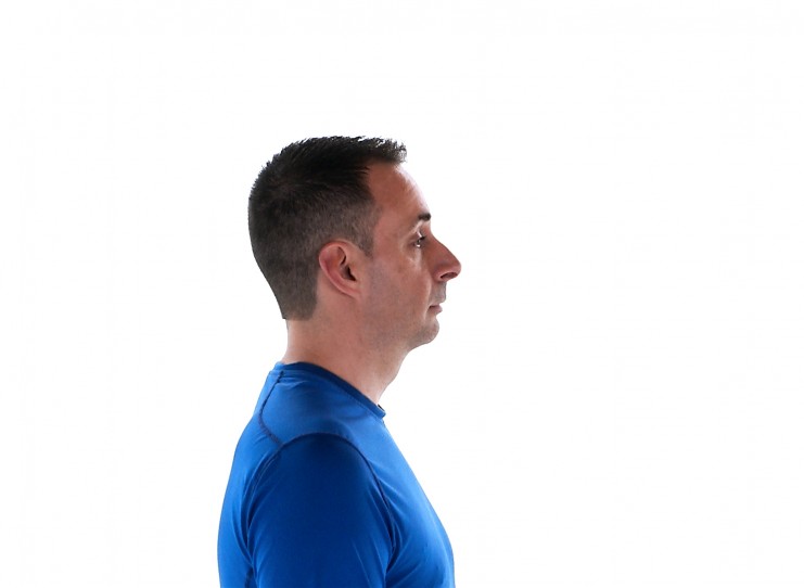
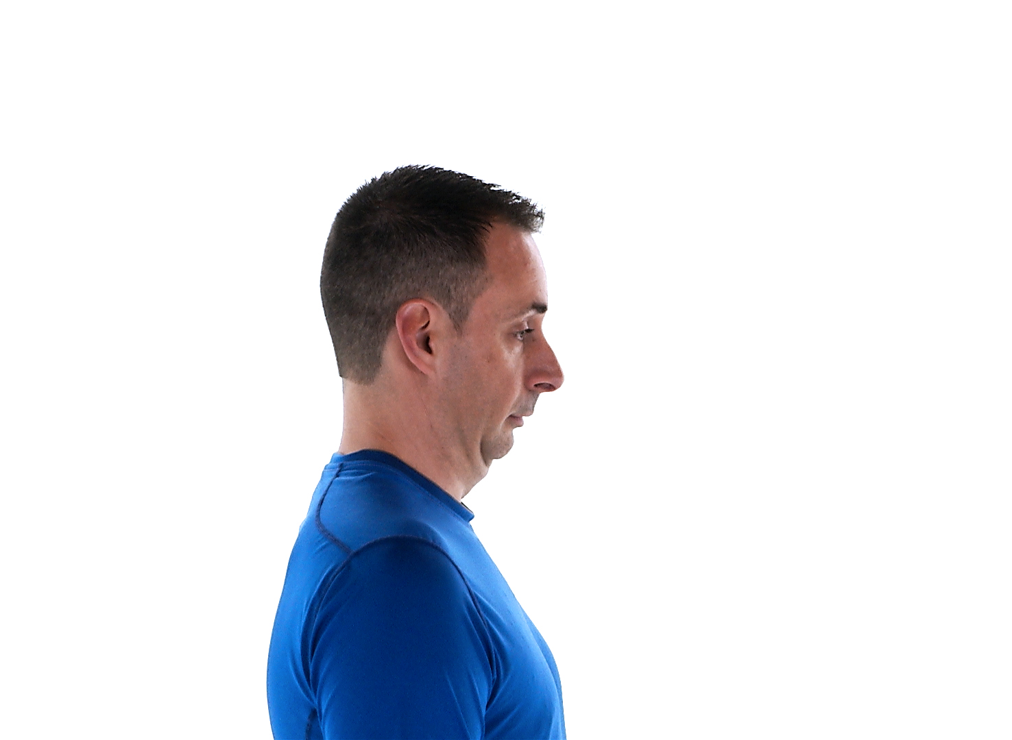
1. Chin tuck
Sit up straight in a chair and look directly ahead of you.
Tuck your chin in without tilting your head down.
Return your head to the original position.
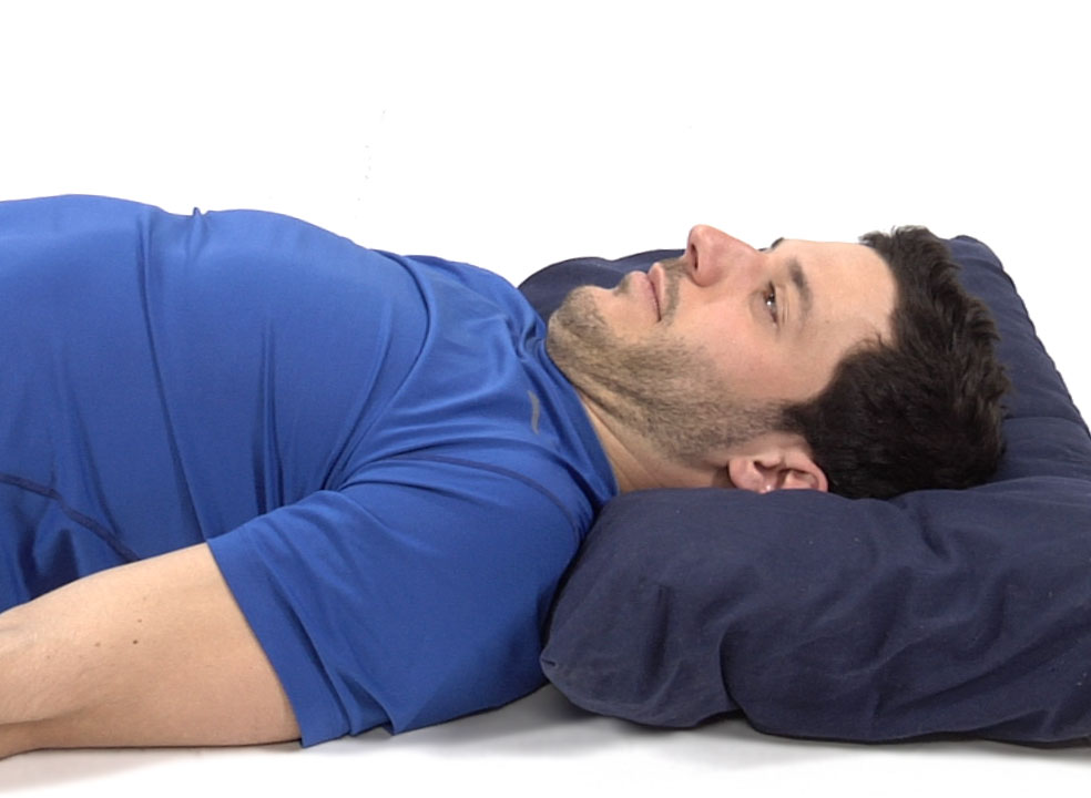
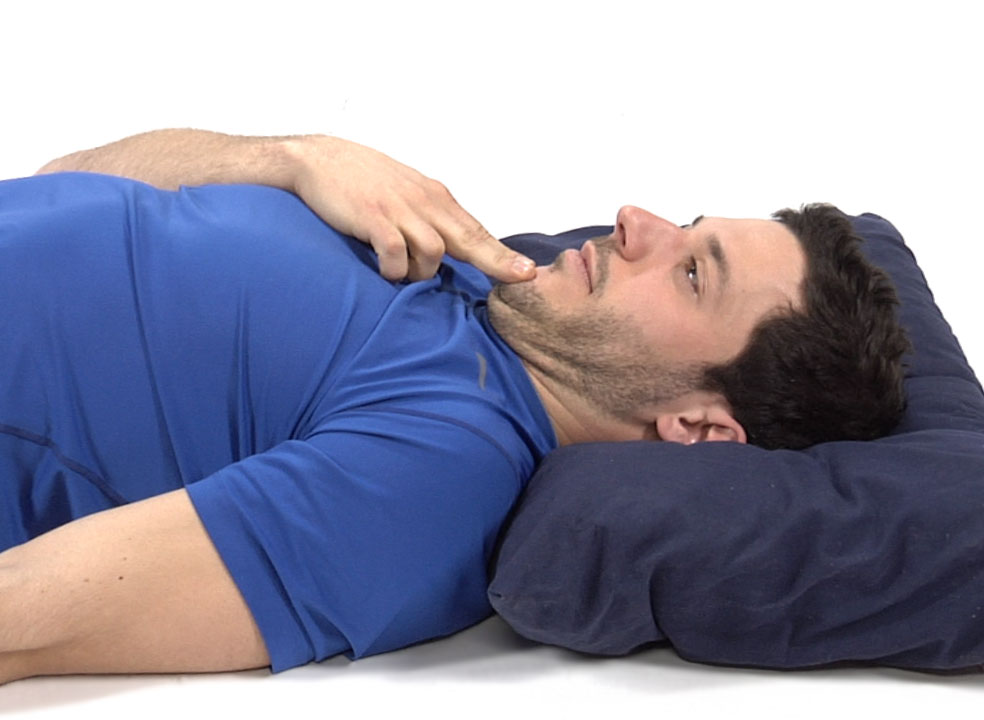
2. Cervical retraction
Lie down on your back with your head resting on a pillow.
Retract your head, pressing down evenly (no tilting up or down) on the pillow with the back of your head.
With your hand, push the chin farther back to feel more of a stretch.
3. Cervical rotation stretch
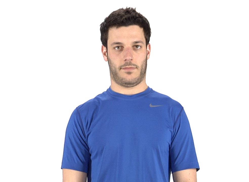
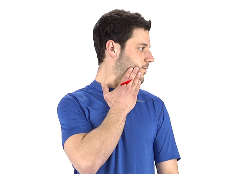
Sit tall with a good posture and a neutral spine (shoulders back, chest lifted, no forward head posture).
Tuck your chin and rotate the head and as the pain subsides, apply overpressure with your hand.
4. Cervical extension + rotation
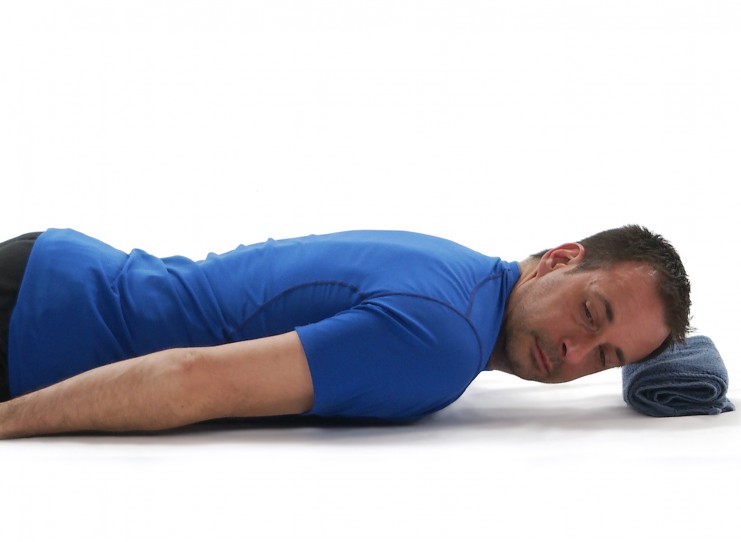
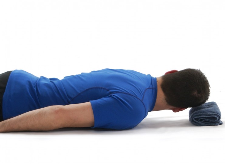
Lie on your stomach with your arms at your side. Place a rolled towel under your forehead.
Tuck your chin slightly to elongate your neck.
Take your head off the towel and rotate it to one side.
Turn to the other side.
Avoid effort/strain sensations between the shoulder blades.
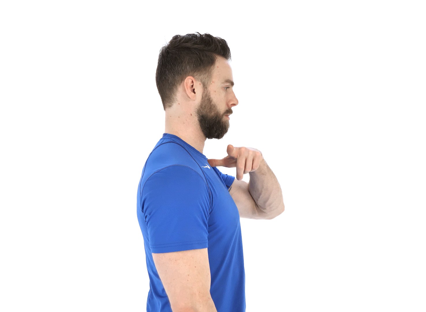
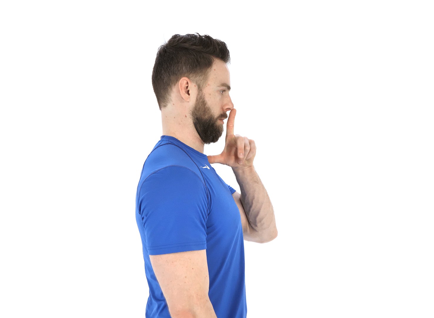
5. Seated trigger finger check for postural correction
Point your finger with your thumb sticking out and resting at the base of your throat above your sternum.
Rotate your hand so the finger points directly up to the ceiling.
Tuck you chin to make way for the finger. You finger should rest at the top of your nose or over your lips depending on your face shape.
This is the corrected head posture.
